Research Paper
Temporal and Regional Circular RNA Profiling in a Tauopathy Mouse Model: Implications for Tau Pathology and Neurodegeneration
Authors
Mohammad Shahadat Hossain1 and Domenico Praticò,2,*
1Department of Neural Sciences, Lewis Katz School of Medicine, Temple University, Philadelphia, PA, USA
2PraticoLab, Philadelphia, PA, USA
*Corresponding author: praticolabalzheimer@gmail.com
DOI:https://doi.org/10.59368/agingbio.20260046
Received: 7/9/2026, Revised: 9/4/2026, Accepted: 9/28/2026, Published: 11/12/2026
Full Article | PDF | Supplementary
Abstract
MicroRNAs (miRNA) are noncoding RNA that regulate gene expression posttranscriptionally and play an important role in neurodegeneration. Circular RNAs (circRNAs) are single-stranded, covalently closed loop RNA molecules recognized as upstream regulators of miRNA. Previous studies have shown that circRNAs are dysregulated in Alzheimer’s and other neurodegenerative diseases. However, a systematic, age- and region-specific circRNA atlas in primary tauopathy is lacking.
To address this gap, we performed comprehensive circRNA sequencing of hippocampal and cortical tissues from a model of human tauopathy, h-Tau mice, at 3, 6, and 12 months of age. Using exploratory differential expression analyses, candidate prioritization across age groups, functional enrichment, and predicted circRNA–miRNA–mRNA network analyses, we prioritized four circRNAs: circ0033491 (Sez6l) and circ0013866 (Abcc1) in the cortex, and circ0004838 (Acaca) and circ0011361 (Ppp2r2a) in the hippocampus. Predicted target networks associated with these circRNAs were represented in pathways related to tau phosphorylation, synaptic function, proteostasis, metabolism, neuroinflammation, and RNA processing.
Together, these findings provide region-specific circRNA profiles for the human tau mouse model and prioritize candidate circRNA-associated networks for future investigation. Although based on computational predictions and exploratory analyses, the identified circRNAs and associated pathways provide a framework for future mechanistic and functional studies of tauopathy.
Introduction
Tauopathies, including Alzheimer’s disease (AD), progressive supranuclear palsy (PSP), corticobasal degeneration (CBD), and Pick’s disease, are disorders characterized by intraneuronal accumulation of hyperphosphorylated tau protein1,2. Tau normally stabilizes axonal microtubules, but its aberrant phosphorylation by kinases such as GSK3b, ERK1/2, JNK, and p38 MAPK at multiple epitopes (Thr181, Thr231, Ser202, Ser396, and Ser422) causes its detachment, somatodendritic mislocalization, and aggregation into insoluble filaments that then become neurofibrillary tangles3–6. Tau pathology propagates in a circuit-dependent manner, originating in the entorhinal cortex and hippocampal CA1 before advancing outward through interconnected cortical networks as defined by the Braak staging framework7,8, with the hippocampus and cerebral cortex representing the two regions of greatest vulnerability.
Noncoding RNAs have emerged as critical posttranscriptional regulators of gene expression in the brain, with microRNAs (miRNAs), short ∼22-nucleotide regulatory RNAs that suppress target mRNA translation or promote degradation9, playing an important role in neurodegeneration. Dysregulated miRNA profiles have been documented in AD brain tissue and biofluids10–13. Our group previously profiled miRNA expressions in the cortex and hippocampus of a mouse model of tauopathy, h-Tau mice, across disease progression, revealing distinct region- and age-specific miRNA signatures14.
Circular RNAs (circRNAs), covalently closed RNA molecules generated by back-splicing of pre-mRNA exons15, are now recognized as major upstream regulators of miRNA. Their closed-loop architecture confers exceptional metabolic stability in postmitotic neurons, and the brain expresses thousands of circRNA species in a cell type-specific fashion3,16. Critically, circRNAs function as competitive endogenous RNA (ceRNA) sponges, sequestering miRNAs through tandem miRNA response elements and thereby de-repressing the cognate target mRNAs17–19.
Dysregulated circRNA profiles have been described in AD cortex20 and Parkinson’s disease substantia nigra21,22, but a systematic, age- and region-specific circRNA atlas in primary tauopathy, a condition where tau is the only pathologic feature, is lacking. To address this gap, we performed comprehensive circRNA sequencing of hippocampal and cortical tissues from h-Tau mice, a model of primary tauopathy, at 3, 6, and 12 months of age. We identified circRNA–miRNA sponging networks that regulate key disease-associated pathways, including tau phosphorylation and dephosphorylation23, epigenetic modifications24, glutamatergic and GABAergic synapses, and microglial efferocytosis25.
Our findings support the novel hypothesis that specific circRNAs have the potential to be used not only as biomarkers but also as therapeutic targets against tau-driven neurodegeneration.
Materials and Methods
Animals and tissue collection
Human tau (hTau) transgenic mice (JAX #005491) were used in this study. Animals were grouped into three age cohorts (3, 6, and 12 months; n = 4 per group, including 2 males and 2 females). Mice were housed under standard laboratory conditions with ad libitum access to food and water on a 12 h light/dark cycle. All procedures were approved by the Temple University Institutional Animal Care and Use Committee (IACUC) and conducted in accordance with National Institutes of Health guidelines. Following euthanasia by CO₂ inhalation, mice were perfused with ice-cold phosphate-buffered saline. Brains were rapidly removed, and the hippocampus and cerebral cortex were dissected on ice, snap-frozen on dry ice, stored at −80 °C, and shipped to Novogene for RNA isolation, circRNA sequencing, and primary bioinformatics processing.
RNA isolation, library preparation, and sequencing
Total RNA was isolated by Novogene using standard protocols for circRNA sequencing. RNA concentration was measured using a Qubit Fluorometer, RNA integrity was assessed using the Agilent 5400 system, and sample quality was further evaluated by agarose gel electrophoresis. All samples met quality-control criteria for library preparation (RIN range: 7.3–9.0).
circRNA libraries were generated following rRNA depletion and RNase R treatment to enrich circRNAs. Libraries were prepared using strand-specific protocols involving RNA fragmentation, cDNA synthesis, end repair, adapter ligation, PCR amplification, and purification. Library quality was assessed using Qubit, insert-size analysis, and qRT-PCR. Sequencing was performed on an Illumina platform to generate paired-end reads.
Read processing, circRNA identification, and differential expression analysis
Raw reads were processed using FASTQ (v0.23.1) to remove adapter-contaminated reads, reads containing >10% ambiguous nucleotides and reads with >50% low-quality bases (Phred score <5). Clean reads were aligned to the reference genome using HISAT2 (v2.2.1).
circRNAs were identified using both Find_circ v1.2 and CIRI2 v2.0.6 and quantified using back-splice junction (BSJ) read counts and reported as transcripts per million (TPM) for expression profiling and visualization. We acknowledge that TPM normalization has limitations for circRNA analyses because BSJ reads do not represent full-length transcripts, and circRNAs do not fully conform to the transcript-length assumptions underlying conventional TPM calculations. Differential expression analyses were performed using DESeq2 v1.42.0 on count-based data. P-values were adjusted using the Benjamini–Hochberg false discovery rate (BH-FDR) procedure separately for each comparison.
The following comparisons were analyzed: cortex (3M vs. 6M), cortex (3M vs. 12M), hippocampus (3M vs. 6M), and hippocampus (3M vs. 12M). No circRNAs remained significant after BH-FDR correction. Therefore, subsequent analyses were conducted as an exploratory candidate-prioritization approach using nominal p < 0.05 and |log₂FC| > 2. To reduce false-positive findings, circRNAs were additionally required to meet these criteria in both age comparisons within the same brain region. Complete differential expression results, including nominal and BH-adjusted p-values, are provided in Supplementary Information S1.
Candidate circRNA selection
Four circRNAs were prioritized for downstream analyses, including one upregulated and one downregulated circRNA from each brain region. Prioritization was based on expression patterns across age comparisons, magnitude of expression change, and exploratory functional annotation analyses.
circRNA–miRNA–mRNA interaction prediction
circRNA–miRNA interactions were predicted using miRanda, and putative miRNA–mRNA interactions were predicted using miRDB. Predicted interactions were incorporated into downstream enrichment analyses and network visualization using Cytoscape v3.10.4.
Functional enrichment analysis
Functional enrichment analyses of circRNA host genes and predicted miRNA target genes were performed using DAVID Bioinformatics Resources. Gene Ontology (GO) Biological Process (BP), Molecular Function (MF), Cellular Component (CC), and Kyoto Encyclopedia of Genes and Genomes (KEGG) pathway analyses were conducted. DAVID uses Fisher’s exact test for enrichment and provides Benjamini–Hochberg-adjusted p-values.
Because of the small size of the input gene sets, no GO terms or KEGG pathways remained significant following BH-FDR correction. Consequently, enrichment analyses were treated as exploratory and reported using nominal p < 0.05. Both nominal and BH-adjusted p-values are provided in Supplementary Information S2–S5.
Region- and age-dependent circRNA expression patterns in tauopathy
To characterize circRNA expression patterns across brain regions and ages, we performed unsupervised hierarchical clustering of all detected circRNAs from the cortex and hippocampus at 3, 6, and 12 months. The resulting heatmap (
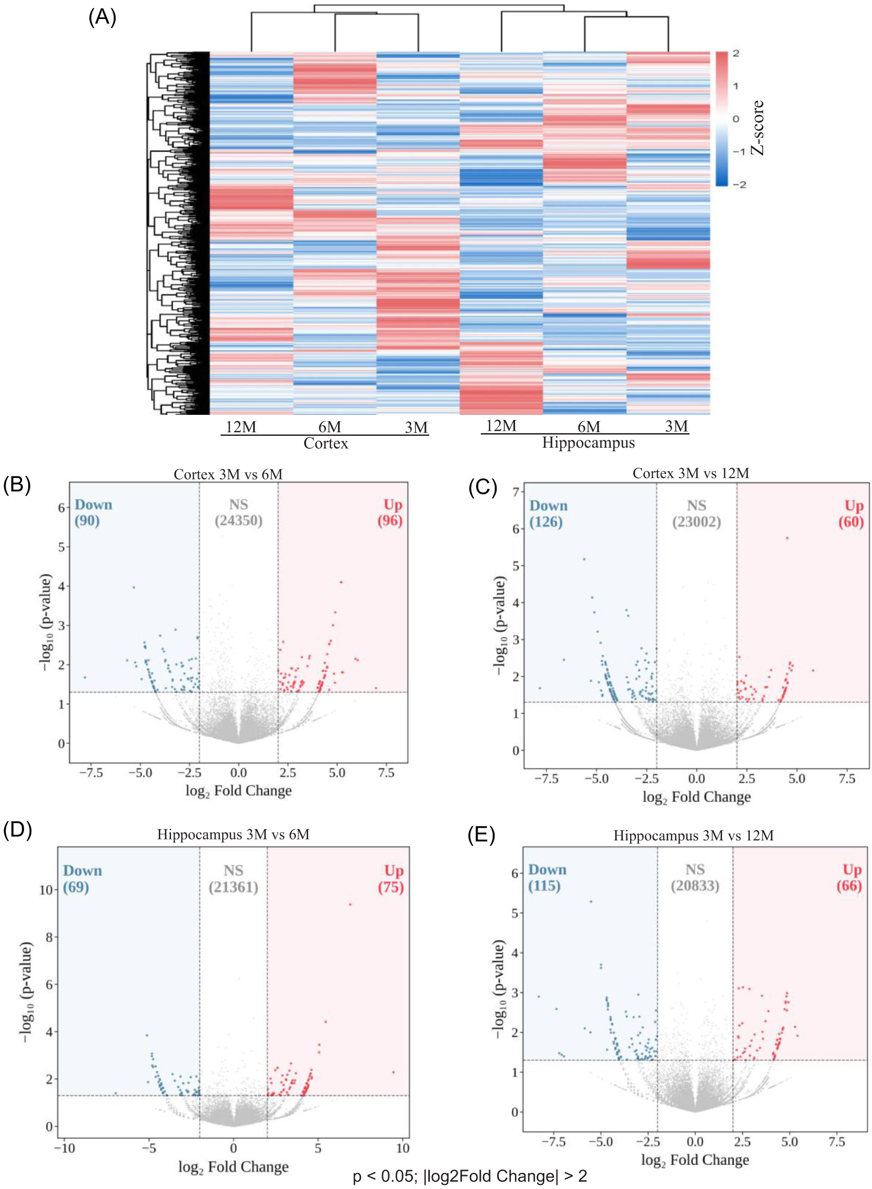
(A) Hierarchical clustering heatmap showing circRNA expression profiles in the cortex and hippocampus at 3, 6, and 12 months (3M, 6M, and 12M). Rows represent circRNAs, and columns represent sample groups. Colors indicate z-score-normalized expression values (red, higher expression; blue, lower expression). Volcano plots showing circRNAs meeting exploratory selection criteria (p < 0.05 and |log₂FC| > 2). (B) Cortex, 3M versus 6M: increased expression (n = 96), decreased expression (n = 90), and unchanged (n = 24,350). (C) Cortex, 3M versus 12M: increased expression (n = 60), decreased expression (n = 126), and unchanged (n = 23,002). (D) Hippocampus, 3M versus 6M: increased expression (n = 75), decreased expression (n = 69), and unchanged (n = 21,361). (E) Hippocampus, 3M versus 12M: increased expression (n = 66), decreased expression (n = 115), and unchanged (n = 20,833). Red points indicate circRNAs with increased expression, blue points indicate circRNAs with decreased expression, and gray points indicate circRNAs not meeting the selection criteria. No circRNAs remained significant following BH-FDR correction.
Because no circRNAs remained significant following BH-FDR correction at the current sample size (see the Methods section), candidate circRNAs were prioritized using exploratory thresholds of p < 0.05 and |log₂FC| > 2 (
Identification of candidate circRNAs
To identify circRNAs showing expression changes at both 6M and 12M, circRNAs meeting the exploratory selection criteria (p < 0.05 and |log₂FC| > 2) in the 3M versus 6M and 3M versus 12M comparisons were intersected using Venn analysis (
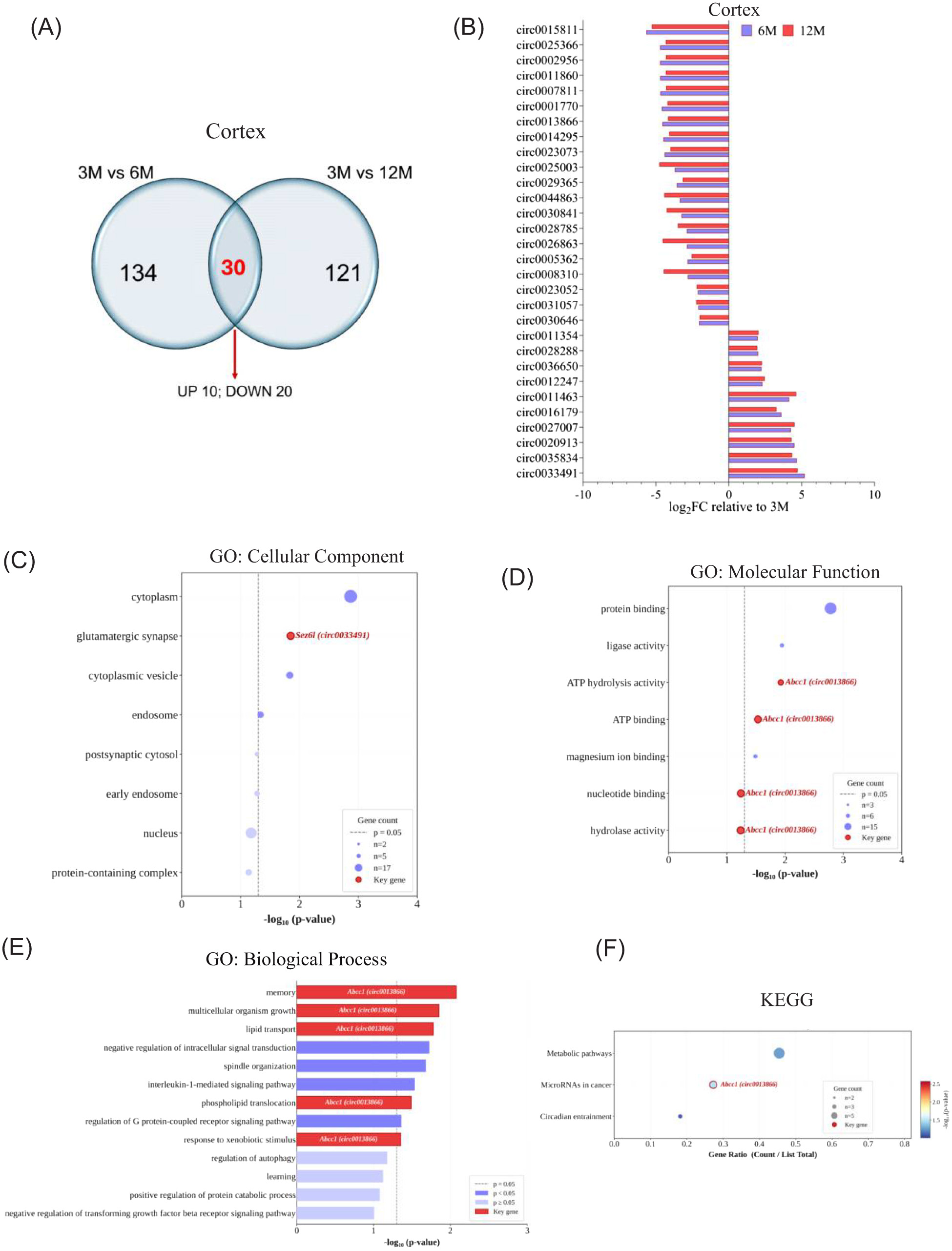
(A) Venn diagram showing overlap between circRNAs meeting exploratory selection criteria in the cortex at 3M versus 6M (n = 134) and 3M versus 12M (n = 121). Thirty circRNAs were shared between comparisons, including 10 with increased expression and 20 with decreased expression. (B) Log₂ fold-change values relative to 3M for the shared cortical circRNAs at 6M (blue) and 12M (red). (C–F) Exploratory GO CC (C), GO MF (D), GO BP (E), and KEGG pathway (F) enrichment analyses of parent genes associated with the shared cortical circRNAs. Dot size represents gene count, and color indicates −log₁₀(p-value). Selected genes are annotated. Dashed lines indicate p = 0.05. No GO or KEGG terms remained significant following BH-FDR correction.
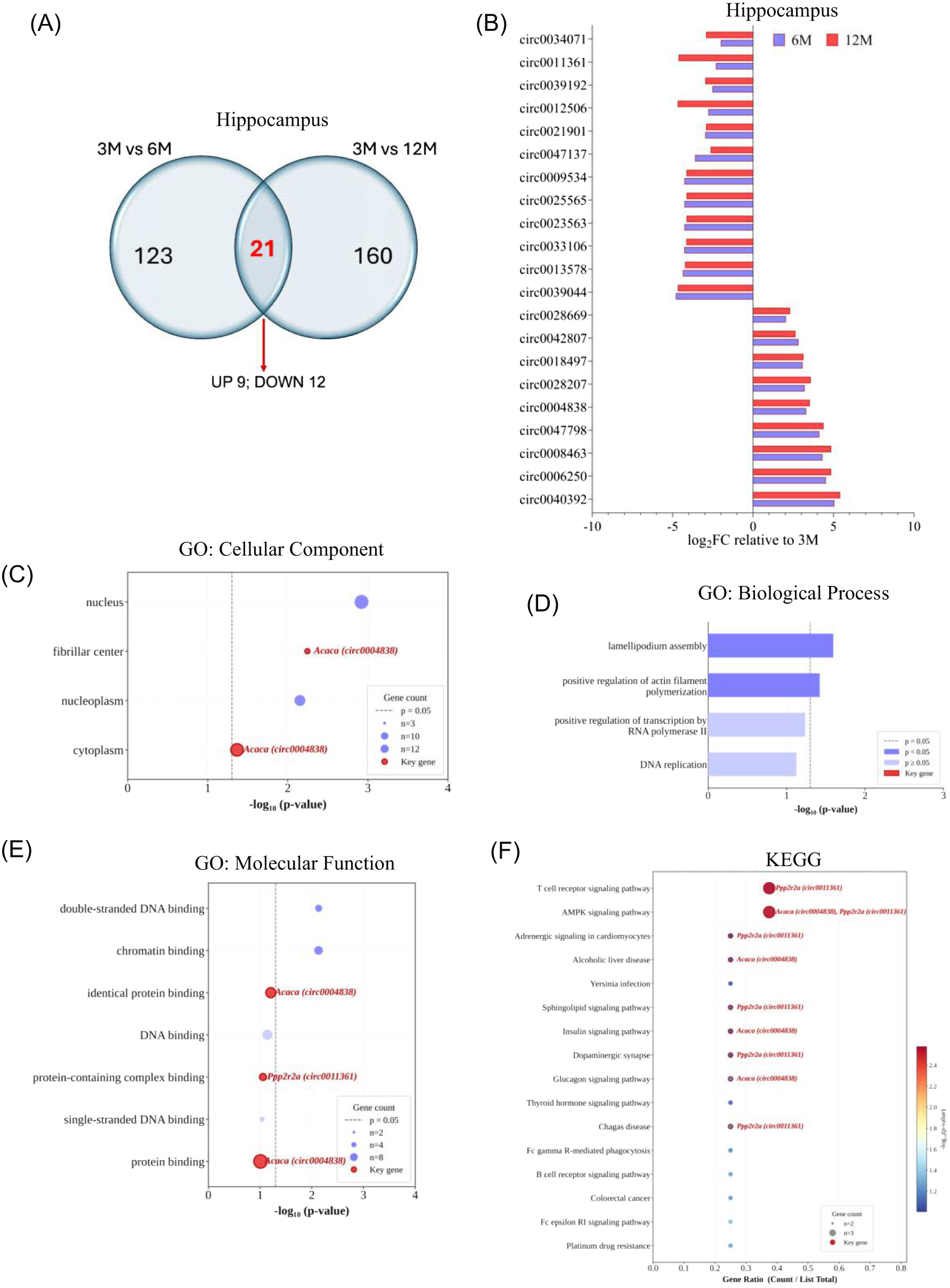
(A) Venn diagram showing overlap between circRNAs meeting exploratory selection criteria in the hippocampus at 3M versus 6M (n = 123) and 3M versus 12M (n = 160). Twenty-one circRNAs were shared between comparisons, including 9 with increased expression and 12 with decreased expression. (B) Log₂ fold-change values relative to 3M for the shared hippocampal circRNAs at 6M (blue) and 12M (red). (C–F) Exploratory GO CC (C), GO BP (D), GO MF (E), and KEGG pathway (F) enrichment analyses of parent genes associated with the shared hippocampal circRNAs. Dot size represents gene count, and color indicates −log10(p-value). Selected genes are annotated. Dashed lines indicate p = 0.05. No GO or KEGG terms remained significant following BH-FDR correction.
Exploratory enrichment analyses of the parent genes associated with the 30 cortical circRNAs highlighted cytoplasm and glutamatergic synapse categories, with Sez6l represented in the latter (Supplementary Information S6 and S7). Additional enriched terms included protein binding, ATP hydrolysis activity, memory, lipid transport, multicellular organism growth, and regulation of G protein-coupled receptor signaling (
Exploratory enrichment analyses of the parent genes associated with the 21 hippocampal circRNAs highlighted nucleus and fibrillar center categories, with Acaca represented in both (Supplementary Information S6 and S8). Additional enriched terms included lamellipodium assembly, actin filament polymerization, double-stranded DNA binding, chromatin binding, and protein-containing complex binding (
Based on expression patterns across time points, magnitude of expression change, and exploratory enrichment findings, four circRNAs were prioritized for further investigation. Among circRNAs showing increased expression, circ0033491 (Sez6l) exhibited log₂FC values of 5.21 and 4.72 at 6M and 12M, respectively, whereas circ0004838 (Acaca) exhibited log₂FC values of 3.31 and 3.53. Among circRNAs showing decreased expression, circ0013866 (Abcc1) exhibited log₂FC values of −4.56 and −4.17, whereas circ0011361 (Ppp2r2a) exhibited log₂FC values of −2.32 and −4.63 at 6M and 12M, respectively (
Prediction of circRNA–miRNA–mRNA networks
To investigate potential regulatory interactions involving the four prioritized circRNAs, miRNA binding sites were predicted for circ0033491 (Sez6l) and circ0013866 (Abcc1) in the cortex and circ0004838 (Acaca) and circ0011361 (Ppp2r2a) in the hippocampus. Five miRNAs per circRNA were selected based on binding score, minimum free energy, and the presence of multiple predicted binding sites (
| circRNA | miRNA | Total Energy (kcal/mol) | Highest Binding Score | Lowest Energy (kcal/mol) | Binding Position(s) | miRNA Length | circRNA Length |
|---|---|---|---|---|---|---|---|
| circ0033491 (Sez6l) | miR-665-5p | −73.50 | 165 | −32.36 | 232, 532, 400 | 26 | 697 |
| miR-709 | −60.91 | 176 | −37.61 | 40, 470 | 19 | ||
| miR-6538 | −57.43 | 168 | −35.27 | 432, 399 | 19 | ||
| miR-3091-3p | −36.05 | 160 | −18.68 | 403, 535 | 22 | ||
| miR-686 | −35.04 | 160 | −19.54 | 204, 116 | 22 | ||
| circ0013866 (Abcc1) | miR-6902-3p | −39.30 | 159 | −20.30 | 52, 1 | 21 | 441 |
| miR-5624-3p | −35.48 | 160 | −20.31 | 415, 163 | 22 | ||
| miR-6540-5p | −32.39 | 159 | −16.64 | 414, 163 | 22 | ||
| miR-7661-5p | −29.77 | 170 | −18.64 | 168, 209 | 25 | ||
| miR-12188-5p | −25.10 | 159 | −25.10 | 201 | 24 | ||
| circ0004838 (Acaca) | miR-7211-5p | −27.14 | 166 | −27.14 | 290 | 22 | 360 |
| miR-7061-3p | −21.73 | 166 | −21.73 | 295 | 22 | ||
| miR-181a-2-3p | −22.97 | 159 | −22.97 | 39 | 22 | ||
| miR-7094-1-5p | −24.67 | 163 | −24.67 | 334 | 23 | ||
| miR-211-5p | −24.02 | 158 | −24.02 | 294 | 22 | ||
| circ0011361 (Ppp2r2a) | miR-30b-3p | −36.31 | 152 | −20.76 | 358, 597 | 22 | 802 |
| miR-6920-5p | −35.33 | 157 | −20.10 | 735, 512 | 22 | ||
| miR-1911-3p | −25.61 | 160 | −25.61 | 677 | 22 | ||
| miR-6908-5p | −25.52 | 170 | −25.52 | 137 | 22 | ||
| miR-693-3p | −24.33 | 168 | −24.33 | 578 | 23 |
In the cortex, circ0033491 (Sez6l) showed predicted interactions with miR-665-5p (−73.5 kcal/mol; three binding sites), miR-709 (binding score 176; two sites), and miR-6538 (−57.4 kcal/mol; two sites). circ0013866 (Abcc1) showed predicted interactions with miR-6902-3p (−39.3 kcal/mol), miR-5624-3p (−35.5 kcal/mol), and miR-7661-5p (binding score 170), each with two predicted binding sites. In the hippocampus, circ0004838 (Acaca) contained a cluster of predicted miRNA binding sites between positions 290 and 334, including miR-181a-2-3p, whereas circ0011361 (Ppp2r2a) showed predicted interactions with miR-30b-3p (−36.3 kcal/mol; two sites) and miR-6920-5p (−35.3 kcal/mol; two sites).
Putative miRNA–mRNA interactions were predicted using miRDB (Supplementary Information S9). Target genes were primarily selected using a miRDB score threshold of ≥80. For two miRNAs, a score range of 55–70 was additionally used to obtain a sufficient number of targets for downstream analyses. The resulting miRNA–mRNA interactions were used for enrichment analysis and network construction.
The predicted circRNA–miRNA and miRNA–mRNA interactions were integrated to construct a circRNA–miRNA–mRNA network. Cytoscape visualization showed a hierarchical network structure in which each circRNA was connected to five prioritized miRNAs and their predicted downstream mRNA targets (
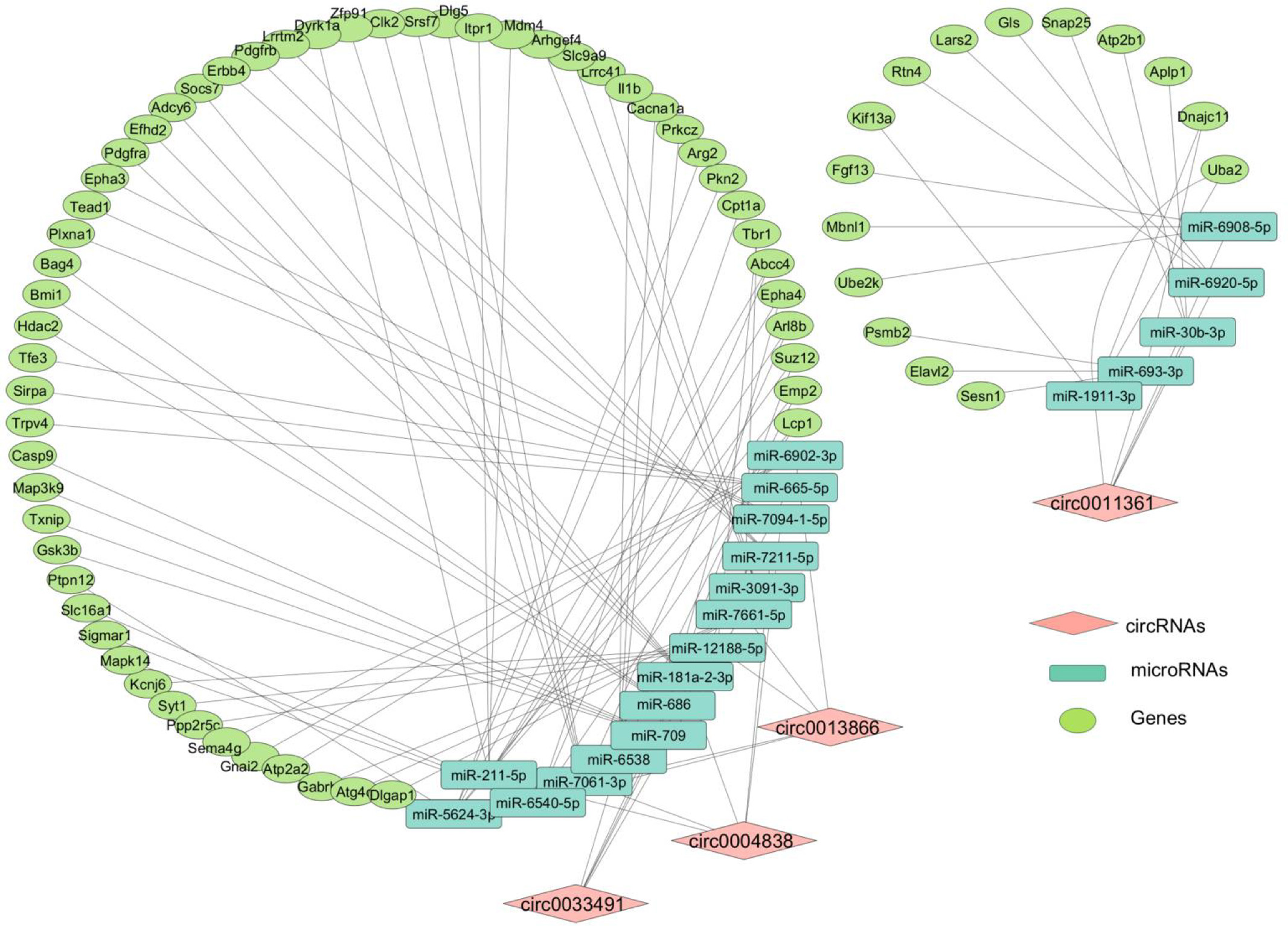
Predicted circRNA–miRNA–mRNA networks for four prioritized circRNAs: circ0004838, circ0033491, circ0013866, and circ0011361. Diamond-shaped nodes represent circRNAs, rectangular nodes represent miRNAs, and elliptical nodes represent predicted mRNA targets. Edges represent predicted circRNA–miRNA and miRNA–mRNA interactions. Networks were generated using miRanda and miRDB predictions and visualized using Cytoscape. All interactions shown are computational predictions and have not been experimentally validated.
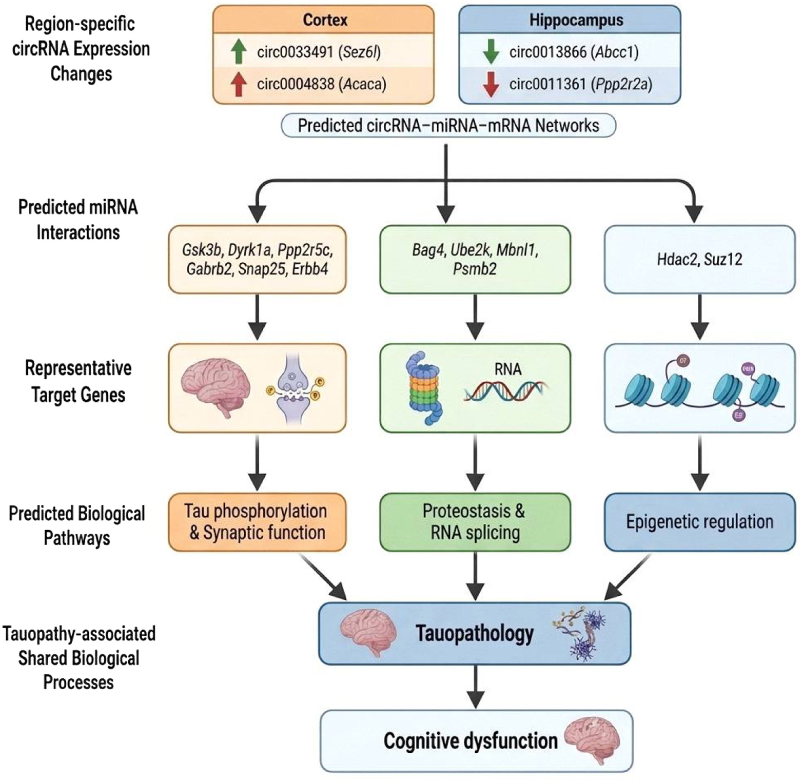
Region-specific circRNAs prioritized from the cortex (circ0033491 and circ0013866) and hippocampus (circ0004838 and circ0011361) were associated with predicted circRNA–miRNA–mRNA networks. Representative target genes are shown as examples of molecules represented within the predicted networks. Exploratory enrichment analyses link these networks to pathways related to tau phosphorylation, synaptic function, proteostasis, RNA processing, metabolic regulation, neuroinflammation, and epigenetic regulation. These pathways were associated with common BPs relevant to tauopathy. This figure represents a hypothesis-generating framework based on computational predictions and exploratory enrichment analyses and does not imply validated mechanistic relationships.
Exploratory functional enrichment analysis
To explore biological pathways represented among the predicted mRNA targets of each circRNA–miRNA network, GO and KEGG enrichment analyses were performed (Supplementary Information S2–S5). Because no GO terms or KEGG pathways remained significant after BH-FDR correction, the enrichment results are presented as exploratory findings and should be interpreted with caution.
Exploratory enrichment analysis of the predicted targets associated with the circ0033491 network highlighted neuronal and synapse-related categories, including growth cone, cell leading edge, glutamatergic synapse, and neuronal cell body. Enriched MFs included ubiquitin protein ligase binding, protein kinase activity, and calcium channel activity. Enriched BPs included regulation of apoptosis, insulin receptor signaling, neuron projection development, calcium ion transport, and ERK signaling. KEGG analysis identified pathways including insulin signaling, MAPK signaling, cell cycle, Hippo signaling, axon guidance, insulin resistance, and AGE-RAGE signaling (Supplementary Information S2).
The circ0004838 network was enriched for synapse-associated and kinase-related categories. Enriched CCs included synapse, glutamatergic synapse, postsynaptic density membrane, postsynaptic density, and GABAergic synapse. Enriched MFs included kinase activity, protein tyrosine kinase activity, and transmembrane receptor protein tyrosine kinase activity. BP categories included protein autophosphorylation, peptidyl-tyrosine phosphorylation, synapse organization, and synapse assembly. KEGG analysis identified gap junction, calcium signaling, cholinergic synapse, and estrogen signaling pathways (Supplementary Information S3).
The circ0013866 network showed enrichment of neuronal, synaptic, phosphatase-related, and apoptosis-associated categories. Enriched CCs included dendrite, axon, glutamatergic synapse, postsynaptic membrane, and endoplasmic reticulum membrane. Enriched MFs included histone deacetylase binding, cysteine-type endopeptidase activity, and protein tyrosine phosphatase activity. Enriched BPs included nervous system development, cellular response to insulin stimulus, apoptotic process, and intracellular signal transduction. KEGG analysis identified efferocytosis, dopaminergic synapse, retrograde endocannabinoid signaling, GABAergic synapse, and morphine addiction pathways (Supplementary Information S4).
The circ0011361 network showed enrichment of synaptic, axonal, and protein-binding categories. Enriched CCs included axon, glutamatergic synapse, synapse, presynaptic membrane, postsynaptic density, and dendritic spine membrane. Enriched MFs included protein binding, metal ion binding, and zinc ion binding. Enriched BPs included nervous system development, positive regulation of neutrophil migration, and axonal fasciculation (Supplementary Information S5).
Discussion
The present study provides a region-specific characterization of circRNA expression across aging in the hTau mouse model and identifies circRNA signatures in the cortex and hippocampus. CircRNA expression profiles clustered primarily by brain region rather than age, consistent with previous reports demonstrating a region- and cell type-specific circRNA expression in the brain26,27. Altered cortical circRNAs were associated with pathways involved in glutamatergic signaling, lipid transport, and memory-related processes, all of which are involved in early synaptic dysfunction and cognitive decline3,28,29. In contrast, hippocampal circRNA dysregulation was associated with nuclear organization, cytoskeletal remodeling, and metabolic signaling pathways, including AMPK-mediated energy homeostasis30,31. Together, these findings highlight region-specific circRNA expression changes during tauopathy progression.
Furthermore, integrative analysis of circRNA expression, predicted miRNA interactions, and predicted mRNA targets prioritized four circRNAs: circ0033491 (Sez6l) and circ0013866 (Abcc1) in the cortex, and circ0004838 (Acaca) and circ0011361 (Ppp2r2a) in the hippocampus. The prioritized circRNA-associated networks included predicted target genes linked to tau phosphorylation (Gsk3b, Map3k9, Ppp2r5c)32–36, synaptic function (Gabrb2, Snap25, Dlgap1, Shisa6)37–40, proteostasis (Bag4, Ube2k, Psmb2, Atg4c)41–43, epigenetic regulation (Hdac2, Suz12)44, and RNA splicing (Clk2, Mbnl1)45. In addition, genes associated with calcium homeostasis and neuronal signaling, including Itpr1 and Atp2b40,46, receptor tyrosine kinase signaling (Dyrk1a, Epha4, Erbb4, Pdgfra)47–49, metabolic regulation (Cpt1a, Slc16a1, Adcy6, Acaca)50,51, and neuroinflammatory pathways (Socs7)52, were also represented within the predicted networks. Although these interactions remain computationally predicted, the representation of multiple pathways previously implicated in tauopathy suggests that altered circRNA expression may be associated with several BPs relevant to disease progression.
Moreover, upregulated circRNAs were associated with kinase-related pathways, including MAPK, insulin, and PI3K-Akt signaling, whereas downregulated circRNAs were linked to phosphatase-related networks involving PP2A-associated regulators. Given the established role of kinase-phosphatase balance in regulating tau phosphorylation53–55, these observations suggest that the prioritized circRNAs may be linked to pathways involved in tau phosphorylation homeostasis. Notably, PP2A-associated regulators were represented within the predicted networks, given that reduced PP2A activity is a well-documented feature of AD56.
Synapses-related pathways were represented across all four prioritized networks. Enriched categories included glutamatergic synapse, postsynaptic density, synaptic transmission, and synapse organization, suggesting that predicted circRNA-associated pathways may be linked to processes involved in neuronal connectivity and signaling. The representation of GABAergic synapse-related pathways within the circ0013866 network is notable given previous reports of GABAergic interneuron dysfunction in AD and related tauopathies57–59. Together, these findings are consistent with the established importance of synaptic dysfunction in neurodegeneration60–62.
Proteostasis-related pathways were also represented across multiple networks. Predicted targets associated with circ0033491 and circ0011361 were enriched for ubiquitin-mediated proteolysis and proteasome-related functions. Representative predicted targets included Bag4, Ube2k, and Psmb2, genes of cellular proteostasis pathways that regulate the balance between proteasomal degradation and autophagy63,64. Given the central role of the ubiquitin-proteasome system (UPS) in tau degradation, alterations in these pathways could influence the accumulation of pathological tau species65,66. Enrichment of autophagy- and lysosome-related pathways within the circ0004838 network suggests a possible involvement of the autophagy-lysosome system. Consistent with this observation, postmortem studies in AD, CBD, and PSP have reported autophagic and lysosomal abnormalities associated with pathological tau accumulation67,68. Similarly, enrichment of pathways associated with histone deacetylation and chromatin remodeling, together with the presence of Hdac2 and Suz12, suggests a possible link between circRNA-associated networks and epigenetic regulation. Notably, Hdac2, a predicted target of the circ0033491/miR-686 axis, has been implicated in the repression of synaptic plasticity genes in AD69, while SUZ12, a core component of the polycomb repressive complex 2 PRC2 complex, mediates H3K27me3-dependent transcriptional silencing and has been linked to age- and disease-associated epigenetic regulation70.
The enrichment of neuroinflammatory pathway components, including TNFa signaling, NF-κB activation through Zfp91, interleukin-1β signaling through Il1β, and efferocytosis across multiple circRNA target networks, suggests an association between these ceRNA networks and neuroinflammatory processes, a well-established feature of tauopathy71–74. Furthermore, the enrichment of efferocytosis-related pathways within the circ0013866 network may indicate involvement of phagocytic clearance mechanisms that have previously been linked to neuroinflammatory responses75–77.
Another notable observation was the representation of RNA splicing and spliceosome-related pathways within the circ0004838 and circ0011361 networks. Predicted targets included Clk2, Srsf7, Mbnl1, Srsf1, and Elavl2, all of which have been associated with alternative splicing regulations. Several of these factors, particularly Clk2, Srsf7, and Mbnl1, have been linked to tau exon 10 splicing and regulation of the 4R:3R tau isoform ratio, a defining molecular feature of multiple primary tauopathies ratio45,78,79. Their representation within the predicted networks suggests a potential association between circRNA expression changes and molecular pathways involved in tau isoform regulation.
Taken together, these observations support a conceptual framework (
Several limitations should be acknowledged. No circRNAs remained significant following BH-FDR correction, and all downstream analyses were therefore based on candidate circRNAs prioritized using exploratory thresholds. In addition, the circRNA–miRNA–mRNA interactions and enrichment analyses were computationally predicted and required experimental validation. The absence of age-matched wild-type controls limits the ability to distinguish tauopathy-associated changes from normal aging effects. Furthermore, the study was designed to evaluate age-associated changes relative to the 3-month baseline; therefore, direct 6M-versus-12M and cortex-versus-hippocampus differential expression analyses were not performed. Future studies incorporating these comparisons may provide additional insight into temporal and regional circRNA expression patterns. The relatively small sample size prevented assessment of sex-specific differences, and the four prioritized circRNAs were not independently validated by qRT-PCR or RNase R resistance assays. Consequently, the circRNAs identified in this study should be considered prioritized candidates for future investigation rather than validated regulators of tau pathology.
In summary, this study provides a region-specific circRNA resource for the hTau mouse model and prioritizes four candidate circRNAs, circ0033491 (Sez6l), circ0013866 (Abcc1), circ0004838 (Acaca), and circ0011361 (Ppp2r2a), for future investigation. Although exploratory, the predicted networks associated with these circRNAs highlight molecular pathways relevant to tauopathy and provide a framework for future mechanistic and functional studies.
Acknowledgments
Dr. Domenico Praticò is the Scott Richards North Star Foundation Chair for Alzheimer's research.
Funding
This research was in part supported by a grant from the National Institute on Health (AG 073622) to D.P.
Author Contributions
M.S.H.: conceptualization, experimental design, data analysis, figure preparation, and manuscript writing. D.P.: supervision, conceptualization, experimental design, conceptual guidance, manuscript review, and editing.
Conflict of Interest
The authors declare that they have no conflict of interests.
Data Availability Statement
The datasets generated and analyzed during this study are available from the corresponding author upon reasonable request.
Ethics Statement
All animal procedures were approved by the IACUC at Temple University and conducted in accordance with National Institutes of Health guidelines for the care and use of laboratory animals.
Supplementary Materials
Supplemental information can be found here: Supplementary.